What is the Importance of Imaging Methods in Dental Treatments?
- 21 Mayıs 2021 07:42


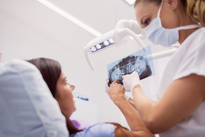
The diagnosis phase is very important for the treatment plan and follow up process to be successful. Successful diagnosis will guide the correct planning. For this purpose, 2D images and three-dimensional tomographs are used.
Where are imaging methods used?
How many types of x-rays are used in the treatment of teeth?
Panoramic x-ray showing the locations of all bones and teeth in the mouth, periapical x-ray showing only two or three teeth together in case of a more detailed idea, bite x-ray showing the left or right part of the mouth used in caries detection; Your dentist will choose one of them according to the application to be made.
Besides all these; There are also three-dimensional imaging methods that may be required before surgical procedures such as implants.
Are the doses of X-rays too high?
The radiation emitted by the radiographs, which play a major role in the planning of dental treatments, contains much less radiation than medical radiographs. For example; like a chest radiograph. Even; Due to the developments in this area, the amount of doses given has been reduced to much lower levels.
How are digital radiographs different from traditional radiographs?
The rate of radiation taken on digital radiography decreases by around 80%. Another important difference is; The recorded records can be stored in computer environment and printed as a dump.
How often should panoramic x-rays be taken?
At what intervals the x-ray will be taken; It varies according to the dental history of the patient. Once a year and twice a year; There are periods that can vary from person to person.
What is the difference between dental radiography and dental tomography?
Dental radiographs provide two-dimensional images. In this imaging method; information on width and height can be obtained; but nothing can be known about the thickness of the tissue.
Dental tomography, especially the tissue thickness that dental radiographs cannot give; They show all the details of tooth and bone tissues. Thus; Possible margin of error in treatment planning is minimized.